Showing posts with label Rohan Bassi. Show all posts

Wednesday, 11 November 2015

Ever Wondered How Alcohol Affects the Nervous System?

Not only in the UK, but all around the world, alcohol is enjoyed by many - drinking is indeed a popular pastime. However, when individuals over-indulge, it can lead to serious health problems including addition even though this is not very common. Nevertheless, in the UK, the NHS estimates that '9% of men and 4% of women show signs of alcohol dependence' (Source: DrinkAware). Over the decades, interest has increased around the effects of alcohol, not only on the physical but also on the mental state. In medicine, researchers will endeavour to find a physiological mechanism to a unexplained mental or behavioural phenomenon. A perfect example of this is the effect of various drugs, illegal and legal, on human behaviour. Quite a few of these substances cause imbalances in the amount of neurotransmitter released from pre-synaptic vesicles into the synaptic cleft in particular areas of the brain. In turn, the frequency of electrical impulses transmitted can fluctuate which can ultimately influence behaviour. To understand this better, it is beneficial to look back in history when the basic physiology and chemistry were uncovered.

Above: Scanning electron micrograph of neurotransmitter-containing vesicles (orange and blue) being released from a pre-synaptic neuron (Source: AnatomyBox)


Pre-1930s, there were noticeable disagreements between academics on how exactly neurons communicate their signals to one another. Was it electrical or was it chemical? It wasn't until 1936, when Sir Henry Dale and Otto Loewi received the Nobel Prize in Physiology or Medicine, that it became clear that these signals were indeed due to chemical transmission. This is possible through the action of neurotransmitters - chemicals which are released into the space between neuron, the synaptic cleft. Some scientists prior to this award did suggest that chemicals were involved, through observing the similarities in nerve stimulation in plants and animals (Source: NobelPrize.org) Loewi managed to illustrate the importance of these chemicals in an elegant way using experiments on frogs. His papers were published in 1921 - these showed that nerve impulses affected the heart using chemical transmission. Firstly, Loewi stimulated the vagus nerve fibres of an isolated frog's heart that had been connected on the other side to a ringer solution. Soon after this, he observed that the strength and frequency of the heartbeat decreased. The fluid remaining was used to surround another frog heart - the vagus nerves were not electrically stimulated. This time, the heart changed it's activity as if it had been electrically stimulated (Source: AnimalResearch.info) It seemed that the fluid has caused this change. Dale's discovery of the action of acteylcholine was inline with Loewi's results and so after subsequent years of research, Dale and Loewi were awarded the Nobel Prize in Physiology or Medicine. 

Chemical transmission is an important concept to understand when we consider how an impulse is able to transmit from neuron to neuron in the brain. It can be understood as a cascade of events beginning with the arrival of an action potential at the axon terminal of the pre-synaptic neuron. The depolarisation stimulates calcium ion channels to open, causing an influx of Ca2+ into the axon. This in turn stimulates vesicles filled with neurotransmitter to migrate to the end of the axon. The vesicles are then able to fuse with the lipid bi-layer membrane to release the neurotransmitter (e.g. acetylcholine) into the synaptic cleft. When these neurotransmitters bind to ligand-gated sodium channels on the post-synaptic neuron, this triggers another action potential to fire. To prevent constant firing of action potentials, a neurotransmitter such as acetylcholine is broken down by an enzyme (in this case, acetylcholinesterase), and the inactive products are reabsorbed by the pre-synaptic neuron through re-uptake transporters. These events will become important when we look at the effect of alcohol on the nervous system.

Above: Schematic diagram showing the transmission of an action potential  (Source: Biological Sciences Review Volume 26, Number 2).

Now, alcohol is one of the few substances than can cross what is referred to in anatomy as the blood-brain barrier (BBB). This is largely a fatty barrier than surrounds the blood vessels in the brain. In medical research, this barrier has been notoriously difficult to overcome when delivering drugs or attempting to treat an array of brain-related diseases, for example Alzheimer's. Until very recently, this has caused problems, however scientists have now found a way of delivering cancer-fighting drugs to targets by breaching the blood-brain barrier. A research team in Canada used 'tiny gas-filled bubbles, injected into the bloodstream of a patient, to punch temporary holes in the blood-brain barrier'. Following this, ultrasound was used to make the bubbles 'vibrate and push their way through, along with chemotherapy drugs' (Source: BBC) This has been a significant breakthrough - I encourage you to read more on the subject here.

The reason that alcohol is able to cross this barrier is that it is lipid soluble. Once alcohol crosses the barrier, it is able to affect the action of neurotransmitters. Before looking at how alcohol comes into play, it is useful to consider the different types of neurotransmitter that exist in the nervous system, and their different modes of action.

Neurotransmitters can either be excitatory (increases the likelihood of an action potential being fired on the post-synaptic neuron) or inhibitory (decreases the likelihood). Examples of excitatory neurotransmitters are glutamate, dopamine and acetylcholine. Glutamate is the most common kind of neurotransmitter in the brain and is thought to be involved in memory and learning. The well-known neurotransmitter dopamine is involved in the mechanisms of motivation and reward. It follows that many addictive drugs utilise the 'feel good' sensation that dopamine causes. And finally, acetylcholine is most commonly used in the contraction of involuntary muscle - it can be released at the site of a neuromuscular junction. A good example of an inhibitory neurotransmitter is gamma-amino butyric acid (GABA) - it's action is known to reduce stress. In fact, about a third of all brain synapses use GABA, and anti-anxiety drugs such as Valium enhance it's action. Another example of an inhibitory neurotransmitter is the amino acid glycine, however this is used mainly in the spinal cord, and is involved in about half of all synapses there, the rest using GABA. (Source: Principles of Anatomy and Physiology 13th edition - G.J. Tortora and B. Derrickson)

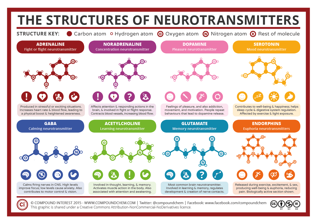
Above: The structure of common neurotransmitters (Source: CompoundChem). See source link for larger image.


Alcohol is widely known as a depressant drug. It is able to decrease excitatory action, and increase inhibitory action. Here, we can look at how alcohol affects the release of GABA. Ethanol increases the amount of GABA neurotransmitter released from pre-synaptic neuron in the brain, by increasing the likelihood that GABA containing vesicles fuse with the bi-layer membrane. To further induce an effect, ethanol encourages GABA to bind more easily to it's corresponding ligand-gated ion channels on the post-synaptic neuron. Subsequently, chloride ions flood into post-synaptic neuron cytoplasm, decreasing the chance that an action potential will fire. In parallel with this effect on GABA transmission, alcohol also affects the action of the excitatory neurotransmitter, glutamate. Ethanol decreases glutamate's excitatory activity, and this effect is quite dramatic considering that glutamate is used in 90% of synapses! (Source: Biological Sciences Review Volume 26, Number 2) The binding of glutamate to it's receptors on the post-synaptic neuron is blocked, thus an action potential cannot be triggered. Since glutamate is used in routes in the brain associated with learning and memory, it is common that people will suffer memory loss after a booze-fuelled night.

The imbalances between inhibitory and excitatory activity do explain the drowsiness, slow reactions and sometimes poor memory people have when drinking a significant amount of alcohol. However one observation we may have forgotten is that to many, drinking alcohol can make them feel good. From research, we know that the reward centre and the pathways associated with it are located in an area of the brain called the striatum. So far, there has been no clear link between alcohol and an effect on the action of the neurotransmitter dopamine. Recall that dopamine is involved in motivation and reward. Where is the link? Well, it is thought that the reward feedback system is usually 'kept in check by GABA inhibition. When this inhibition is suppressed, the reward system becomes more active'. Remember that alcohol does cause suppression of GABA inhibition as it encourages more GABA to bind to the post-synpatic neuron!

Above: Schematic of the brain showing the location of the striatum (yellow-orange region) (Source: Biological Sciences Review Volume 26, Number 2)

To conclude, we can see that in order to understand how alcohol can affect the nervous system, it is important to appreciate the biological cascade of events that occur during chemical transmission at the synapse. Although alcohol can be enjoyed in moderation, the public must be aware of the potential health complications associated, including liver disease, weight gain and sleep disruption. The incidence of liver disease particularly, is rising in the UK. Bear in mind that this disease not only affect adults, but also the young as well. Let us not forget also of the societal problems that can arise due to alcohol abuse, which include antisocial behaviour and violence in extreme cases.

Even today, alcohol still presents unsolved mysteries to researchers. However with continuous advances in technology, medicine, and neuroscience, how the brain is affected by substances is becoming clearer and clearer.


Additional credit: Oliver Freeman is a writer for the Biological Sciences Review and is also studying for a PhD in neuroscience.
 - Michelle Roberts, for her article published on the BBC website, 'Scientists breach brain barrier to treat sick patient'. Read more on the subject here.

Saturday, 7 November 2015

Revolutionary Platinum-based Chemistry: A New Hopeful for Cancer Therapy

As many would agree, one of the most well-known treatments that cancer patients undergo is chemotherapy. Today, it is not unusual for platinum compounds to be used in chemotherapy - cisplatin is a notable example.


Above: False-colour electron micrograph of cancer cells (Source: Wellcome Collection)


In medicine, particularly pharmacology, the shape of molecules are extremely important. In cisplatin, the oxidation state of platinum is +2 and the molecule is said to be square planar. This means that all of the atoms lie in the same plane, forming a square if you were to join up the atoms with imaginary lines. Each of the groups, Cl- (Chloride - with an oxidation state of -1) and NH3 are called ligands, and because each of the different groups are on the same side of the molecule, the platinum compound is said to have a cis structure. In the body, cisplatin's basic chemistry works as follows:

Since, in the bloodstream there is a high concentration of chloride ions, none of the ligands on the molecule are substituted (NHgroups are more resistant to this substitution). However, once inside the cell the environment is very different. In fact, there is a much lower concentration of chloride ions, and the chloride ligands are replaced by water molecules. Now the cisplatin compound is activated. This is a perfect illustration of Le Chatelier's principle:

"If a dynamic equilibrium is disturbed by changing the conditions, the position of equilibrium moves to counteract the change"
(Source: Chemguide)

Above: Displayed formula of cisplatin


Although cisplatin  has proved effective, over the years, several similar drugs such as carboplatin have been produced to maximise efficiency and potency. The drug works by disrupting cell replication, thus leading to cell apoptosis (death). The mechanism for this relies on the substitution of those chloride ligands for water molecules once inside the (cancer) cell. From this point, nitrogen atoms from the nucleotides forming DNA substitute the water molecules. Water molecules tend to be easily replaced. In this situation, cisplatin  is effectively bound to the DNA, causing the nucleotide chain to bend due to formation of a 'kink'. In DNA replication, the shape of DNA is very important and many checks are made by feedback mechanisms to make sure the cell has proceed to divide by mitosis. Once cisplatin is bound, mitosis can no longer take place - cell death results.

However, current research into the field of photoactive compounds has proved promising - the research could be a step to producing cancer drugs that can be activated at the tumour site using lasar technology. Firstly, let us consider the properties of platinum. It has a silverly coloured surface (without tarnish) and is used in catalysis - catalytic converters in road vehicles is an example. So why does this metal prove useful in the body? Platinum is considered a relatively safe, it is said to be biologically compatible due to it's inability to react with body tissue. However, in medicine, what we are more concerned about is whether we can use platinum compounds. In fact, these compounds are used but scientists are aware of the toxicity of such substances. Platinum ions that are bonded to several ligands help to channel potential toxicity in a useful way, often to produce life-saving drugs.

The challenge now is to produce treatments that are even more localised to the cancer cells. This is inevitably the subject of research in universities and pharmaceutical corporations across the globe. Existing cancer drugs work well simply because cancer cells are subject to more damage than normal body cells - cancer cells proliferate at a faster rate. Note however that normal body cells can still become affected. As many may have experienced, the side effects of chemotherapy can be quite extensive, nausea and kidney damage to name a couple. The human body is simply trying to reject the foreign substance introduced, and this is what drives scientists to look for new solutions. 

The key fact to know is that whilst cisplatin and other similar drugs are not tumour specific, how they are activated can be controlled. Platinum, like many other transition metals, have multiple oxidation states. Platinum(IV), Pt4+ complexes 'have been proven to be inactive and non-toxic inside cells, but only in the dark' (Source; Chemistry Review Volume 24, Number 4). One reason for this is to do with the shape of these platinum(IV) complexes. With an oxidation state of +4, platinum is able to form 6 bonds to ligands in an octahedral arrangement. Remember than a platinum(II) compound can only form 4 bonds with ligands in a square planar structure. This higher oxidation state enables the molecule to be less reactive and therefore ligands tend to be become replaced. This is very relevant, recall that cisplatin has it's chloride ion ligands replaced by water molecules once inside the cell. However, it is important to remember that the reduced reactivity in the case of platinum(IV) compounds is true in the dark. These compounds can be photo-activated - light enables the configuration of the molecule to change. This process is irreversible:

Above: The photo-activation of a platinum complex - X and Y denote alternative ligands (Source: Chemistry Review Volume 24, Number 4)

This reaction is able to occur to due to the phenomenon of electron transition. An election which absorbs light energy is able to be promoted to a higher energy state, and therefore a higher energy orbital with an atom. In transition metal chemistry, it is common knowledge that when ligands bond to the central metal ion, this causes the d-orbitals of the metal ion to split. The orbitals are split into two levels, one with a higher energy level than the other. The very fact that these complexes can absorb light energy means that transition metal complexes are often colourful. During the above reaction, electrons in the central metal ion 'jump' to a higher energy level. Any remaining light that is not absorbed is reflected back. These electron transitions can cause multiple changes, such as a change in oxidation state of the central ion, or substitution of ligands. Controlling this activation could indeed be a useful tool in cancer therapy, it could potentially have wider applications in medicine. After activation, the cisplatin-like compound can then perform it's anti-cancer wonders. 

Above: Diagram showing that the energy of a particular wavelength of light is equal to the energy required to promote an election to a higher energy level (Source: Chemguide)


Using these platinum compounds does leave room for flexibility. For example, scientists would be able to change the ligands to vary the amount of light energy absorbed (to cause d-orbitals to split). However, getting this energy quota just right is a challenge, and is still the subject of research. 

So what are the applications in cancer treatment? Any treatments should be as safe as possible, so it is important what type of light should be used to activate the platinum complexes inside the body. Now, most complexes tend to be activated by blue or even UV light, however these frequencies of light do not penetrate tissue as well as red light does. Bear in mind that UV can damage tissue - red light seems a relatively safe option. A compromise must be made as red light would mean that it is less likely that a complex would be activated. Nonetheless, 'some promising Pt4+ complexes have been made, which are non-toxic in the dark but once activated have a high toxicity towards cancer cells'. After all, it is completely dark inside the body, therefore a laser would need to be used to activate the chosen drug. A laser would be a suitable choice due to it's precision - it would be much less likely that a healthy body cell would be affected (Source: Chemistry Review Volume 24, Number 4)

As with any new treatment, this new concept would need to be subject to vigorous testing through a series of clinical trials. Safety and effectiveness are two crucial criteria that will need to be evaluated during the course of these trials in future. 

Additional credit: Louise Tear who wrote an article in the Chemistry Review, which was inspired by an undergraduate research project completed under the guidance of Professor Peter Sadler.
Further credit: Professor Sadler who wrote a short piece for theInformationDaily.com, 'Using precious metals to fight cancer', following research at The University of Warwick. 

Further reading: BBC, 'Chemists create new way to fight drug resistant cancer'.
Macmillan Cancer Support - 'Cisplatin - Cancer Information'

Friday, 6 November 2015

Has This Been Humanity's Deadliest Threat to Date?

Over the course of centuries, humans have witnessed the wrath of many deadly endemic, epidemic and pandemic diseases. Some notable examples include the uprising of small pox and the Bubonic plague. The number of deaths worldwide that have resulted are alarming. However, what is more profound is how fast the pathogens of these diseases spread in a population. In later years, the impact of these epidemics often become the subject of academic study in Medicine, in particular, epidemiology. In addition to these giants of infectious disease, there is another worth mentioning, which could be debated as 'the greatest medical holocaust in history' - the Spanish Flu of 1918.

Above: A Spanish flu ward at Fort Riley, Kansas, in 1918. (Source: The Guardian)

Caused by the H1N1 Influenza virus, the Spanish Flu was capable of rapid transmission, which resulted in it's success - 500 million people infected worldwide (one fifth of the world's population at that time (Source: Census.gov)). The fact that the infection numbers were indeed astronomically large, in the years post-pandemic, it was difficult to make an estimate of the mortality rate. Another reason is that many different countries around the world were affected by a preceding war, and different countries were affected to different extents. However, most sources indicate that the number of deaths ranged between 10-20% of those infected, i.e 50-100 million (Source: Archives.gov - The Deadly Virus). To put this into comparison, just over 17 million were killed over the duration of the Great War (Source: BBC). Despite the magnitude of destruction that the Spanish Flu inflicted, it has become a subject of lesser interest over the years. Looking back at these events, what could we learn to move ourselves forward in the medical field?


One of the great mysteries surrounding the Spanish Flu pandemic is that of the origin of the virus. Some of the latest media report that this virus is likely to have originated from the Far East, in particular, China. However, previous suggestions for the origin location range from Midwest America to France! It is generally accepted that the virus later mutated, causing the most destruction. According to the National Geographic, "new research is placing the flu's emergence in a forgotten episode of WW1: the shipment of Chinese labourers across Canada in sealed train cars." During the War, there was an increasing demand for labour, especially behind the British and French lines.


Above: Public notice for influenza in 1918 (Source: Wikipedia)


Unfortunately those that were infected often suffered unpleasant symptoms: bleeding from the nose and ears was common as well as (after autopsy) swollen hearts and lungs that had become solidified. Some figures showed that some lungs after autopsy measured up to six times their normal weight. The explanation for this is the build up of fluids (oedema) during the course of infection. This accumulation of fluid would have been a significant obstruction and gas exchange would have become increasingly difficult. It follows that as a result of this, many of those infected would die of asphyxiation. One of the physicians working at a military camp near Boston, Massachusetts in September 1918 describes the symptoms of asphyxiation one would typically have in vivid detail:

"Two hours after admission they have mahogany spots all over the cheek bones, and a few hours later you begin to see the cyanosis extending from their ears and spreading all over the face, until it is hard to distinguish the coloured men from the white. It is only a matter of a few hours then until death comes and is is a struggle for air until they suffocate. It is horrible. One can stand it to see one, two or twenty men, but to see these poor devils dropping like flies sort of gets on your nerves." 
                                                 
                                                                         - A physician stationed at Fort Devens, Boston, September 1918 (Source: Voices of the Pandemic) 

As well as these conditions that resulted from infection of the virus, often, many others would become ill from secondary infections such as pneumonia - a bacterial infection. The influenza virus is able to penetrate the respiratory system and damage the cilia and epithelial cells lining the lungs. The immunity of the infected is weakened due to the cells of the immune system losing their function. Thus, one becomes increasingly susceptible to pneumonia. 


(Above: Orginal photograph of the H1N1 virus, taken in the CDC Influenza Laboratory) 

As we know, the Spanish flu was caused by the H1N1 virus. What does this mean? Any virus that contain the letters H and N each followed by a number indicates that the virus is type A influenza. The letters H and N refer to haemagglutinin and neuraminidase respectively, the distinctive membrane proteins on the virus. Haemagglutinin binds to receptors on host cells. This causes fusion of the two membranes and deadly infiltration of the viral content. Neuraminidase acts at the end of the viral replication cycle - it 'cleaves' the new virus from the host cell. Now, the cycle is able to occur again and again, and other neighbouring cells become infected. Moreover, the proteins can actually prove very useful - they are extracted from circulating strains, purified, and use in a flu jab vaccine that is given every year.

However, what made H1N1 in 1918 such a big problem was the concept of genetic drift. This became apparent in 2005, when a group of American scientists sequenced the genome of the 1918 flu virus. The tissue sample came from a female patient who was buried in an Alaskan permafrost. The shift was gradual, initially being carried in an avian host. The H1N1 was able to mutate during the course of the pandemic, making it's infection very potent. A mutation in the genome would have caused the subsequent virus to produce subtly different variations of haemagglutinin and neuraminidase. As a consequence, antibodies produced by the host will no longer be able to bind to these proteins. The virus evades the immune response.


Above: The pathogenesis of an influenza A type virus (Source: Biological Sciences Review Volume 27, Number 4)


You might argue that perhaps only the most vulnerable would have been at risk. However the virus was evidently very potent and not discriminatory it would seem. The flu was prevalent in rural as well as urban areas - even the most remote parts of Alaska were affected! Usually, young adults tend to be the least affected when it comes these types of infectious diseases - their immune systems are generally well developed. However, for the Spanish flu, it was the exact opposite. This group tended to be severely affected, along with the vulnerable groups (elderly and young children). One astonishing statistic is that the average life expectancy of the USA dropped by twelve years during one year of the pandemic alone. (Source: Archives.gov)

Above: Age profile of deaths from Spanish flu (Source: Data from Centers for Disease Control and Prevention)

In the graph above, we can compare the deaths for each age group during the period 1911-1917 to the year 1918 - the year of the Spanish flu. What is unusual is the spike in deaths in the age group for young adults. Over the years, this has intrigued epidemiologists - however one theory that does exist to explain this oddity of flu epidemics is the 'cytokine storm'. This relates to the idea that the young and healthy have the most powerful and effective immune systems. However, during an infection with flu, the immune response can too excessive, becoming detrimental to health. Cytokines are chemical released by cells of the immune system during an infection to provide a means of cell communication. Some cytokines accelerate chemical processes, whilst others inhibit them. They also cause increased inflammation, swelling, and vasopermiability (the blood vessels become more permeable). Usually, this would help to fight the infection, however sometimes this response can come at the expense of an organ that has an oedema (and reduced blood supply). A consequence of this is tissue scarring, and then multiple organ failure. So, in the case of Spanish flu, an 'overreaction' of the immune system can indeed prove fatal (Source: Biological Sciences Review Volume 27, Number 4).


What could be done in the future? According to the World Health Organisation, the next pandemic 'will kill between 2 and 7.4 million people'. H5N1 (bird flu) is considered the most dangerous currently. In future, epidemiologists will need to keep watch for emerging epidemics that could potentially become catastrophic pandemics. In the field of infectious diseases, emphasis is being placed on prevention, more than ever before.

In addition to the reference provided above, credit should be given to Bethany Butcher who wrote an article on Spanish flu for the Biological Sciences Review April issue, 2015.
Extra reading:


Tuesday, 17 February 2015

The Polio Virus - Close To Eradication?

Also known as Poliomyelitis, polio is an infectious disease, caused by the poliovirus. Infection can result in infantile paralysis and muscle spasms in its worse cases. The virus can be ingested through contaminated food or water, and be absorbed through the gut wall. From here it is able to move to the spinal cord and paralyse its nerves.

Above: Scanning Electron Micrograph of the poliovirus (Source: Polioeradication.org)

Typically, polio is relatively prominent in developing countries, Asia containing most of the transmission. Evidence suggests that the poliovirus has been around for a very long time, some sources quoting that it infected people in the prehistoric ages. However up until the 20th century, major epidemics were simply unknown. Until this time, the virus spread was known to be endemic - this means that in an area, the virus' transmission remained fairly constant, with no real surges in infection. In the 20th century however, Europe, followed by the Americas began to experience widespread epidemic of the disease. What is interesting is that the disease saw its highest incidence in the summer months of each year.

'At its peak in the 1940s and 1950s, polio would paralyse or kill over half a million people worldwide every year' (Wikipedia).  Franklin D. Roosevelt was one of the most notable people to be infected with the poliovirus, becoming permanently paralysed from the waist and down. (However this seems quite an unusual case as the poliovirus is typically known to induce infantile paralysis. Therefore, there is debate surrounding this specific case) This is a staggering number, considering that the rates of transmission were 'controlled' only couple of decades before this period. These rather catastrophic events initiated a global medical response, with research funding increasing dramatically.

Probably one of the most known national crises with polio was the Copenhagen polio epidemic in 1952. However epidemics of equal magnitude were spreading across America at the time.
"Anyone wandering in to Ward 19 of Copenhagen's Blegdas Hospital in the autumn of 1952 would have been confronted by an extraordinary sight. In each of the seventy beds arranged in two straight lines lay a child paralysed with polio with a hollow plastic tube inserted into the trachea through a cut in the neck - a tracheostomy - to which was attached another long piece of tubing, at the end of which was a rubber bag. Next to each bed sat a young medical student who, every few seconds, would squeeze the bag, blowing oxygen through the tubing into the child's lungs and then letting go, repeating this action for six hours at a stretch[...]According to Ann Isberg, one of the children, 'it was not a sad time', rather 'like [during the] war there was a spirit of resistance - everybody was doing their best'."
 - An account describing the atmosphere in a hospital ward, housing polio patients, from 'The Rise and Fall of Modern Medicine' written by James Le Fanu, M.D. 
Special respiratory centres were then established in the Blegdas Hospital, under the initial influence of anaesthetist Bjørn Ibsen, in order to help ventilate the polio patients (whose breathing was compromised by paralysis). This was essentially the birth of intensive care. Following these events, which constituted several 'summer plagues', in 1957, Jonas Salk, an American researcher helped synthesise the first successful polio vaccine. Choosing not to patent it, he allowed the vaccine to be distributed all over the world for free. This led to a vast decline in polio cases. By the late 1980s, many described the virus 'being close to eradication' in most countries.


Above: Polio ward in Hynes Memorial Hospital in Boston, 1955 (Source: Dailymail)

Above: An Iron Lung machine, used in the assistance of ventilation/breathing in polio patients (Source: Centers for Disease Control and Prevention's Public Health Image Library (PHIL) - via Wikipedia


Here is one staggering statistic: 'Polio cases have decreased by over 99% since 1988, from an 
estimated 350 000 cases then, to 416 reported cases in 2013' (The World Health Organisation Polio Fact Sheet)

Despite this, new cases of polio have been emerging in Afghanistan, Pakistan and Nigeria. In 2014, the World Health Organisation saw that this surge in cases meant the declaration of a public health emergency of international concern (PHEIC). The vital key to eradicating polio completely is nationwide herd vaccinations, as the virus cannot survive for long outside a host.


The Final Stages in Eradicating Polio

As of early 2015, scientists have been collaborating internationally in order to develop a synthetic vaccine in the hope of eradicating the last remaining strain of the poliovirus. Together, the World Health Organisation and the Bill & Melinda Gates Foundation have provided a $674,000 (£438,000) grant to fund the research. The main problem with current vaccines is that because they utilise the weakened (attenuated) form of the virus, in some patients, this may initiate an immune response. This would mean that the virus can be 'reactivated' and passed on to those that have not been vaccinated. However a synthetic vaccine means no genetic material which essentially indicates that the vaccine contains no virus.


We are extremely close to eradicating the poliovirus off the face of the Earth. What a human feat that would be.


References

In addition to sources given above:
- Credit to the World Health Organisation for their information and data on the poliovirus and the polio vaccinations
- Additional Credit to BBC Science Correspondent, Jonathan Amos for his article on the new synthetic vaccine for polio, 'Synthetic vaccine sought to finally eradicate polio' (14th February 2015) Read more.
- Additional Credit to James Le Fanu's 'The Rise and Fall of Modern Medicine' which features the events surrounding the Copenhagen polio epidemic and the birth of intensive care.


Sunday, 15 February 2015

Hepatitis C - Can We Use Genetics To Find Effective Treatments?

As devastating as viral infections go, Hepatitis C is one of the most severe, in worst cases leading to cirrhosis - liver tissue scarring. There have been cases where individuals have developed cancer as a result of the destruction due to the Hepatitis C virus. The success of the Hepatitis C virus is largely due to the survival of its most deadliest strains. The fact that humans are the only species as far as we know that can be infected with HCV makes us somewhat more vulnerable. What is more interesting is that despite all our current knowledge on viral replication and adaptation, the mechanism of this virus infiltrating cells and causing damage is not fully understood. You could argue this 'void' in our scientific knowledge is preventing us from devising effective treatments, not only to this particular type of infection, but others too. However just like any other virus strain, the Hepatitis C strain which causes disease does utilise the host cells 'machinery' in order to replicate itself multiple times. It follows that whole new virions are produced from the newly synthesised viral protein. Despite there being some individuals who are able to fight off the virus, most people develop a chronic infection which can last as long as a lifetime. Continued degrading of the liver over a prolonged period means that eventually the liver cells lose function -  only a liver transplant can save the life of the patient.

What seems reassuring statistically, is that according to the World Health Organisation, around 3% of people worldwide are affected severely by the Hepatitis C virus (HCV). A small percentage one may consider, however it equates to a very large quantity. Additionally, a large number of people may have not been diagnosed with the infection as the infection takes time to develop. This can only mean that the actual number of people affected chronically with HCV is much higher.

A Summary of HCV Transmission:

A blood-bourne infection, HCV is commonly transmitted through (vascular) medical operations, or through intravenous drug usage. There have been nationwide catastrophes in some countries where blood, contaminated by HCV, has been used for transfusions. The identification of the virus was too late, and many people were medically affected.

The origin of the spread of HCV is largely unknown, although many agree that it could have been initially spread by some kind of vector, a mosquito being a predictable example. It has been discovered that the virus can spread through sexual transmission, however blood-to-blood contact is seen as the most efficient method of the virus' transmission.

Above: Electron Micrograph of the Hepatits C Virus (HCV) isolated from cell culture (Wikipedia)


The Significance of the Viral Genome:

The HCV, as characteristic of many viruses, is able to mutate at a frequent rate, making it very difficult to develop a long-lasting vaccine. The length of its genome is relatively short at around 10000 nucleotides long. The primary reason for the rapid mutating of the HCV genome, is that unlike in humans, the HCV virus has no mechanisms in place to proof-read its own DNA. This frequency results in copying errors during the process of DNA replication. Combining this feature (or rather a lack of it), with HCV's ability to replicate at an extraordinary rate, results in many mutations in the viral DNA sequence.

To most people, a mutation is seen an event detrimental to the host organisms, however this isn't always the case. In viruses, due to the large number of mutations that occur, mutations can sometimes be advantageous. Therefore the virus is able to acquire a protein that serves valuable to virus, allowing it to survive in more extreme conditions. One application of this is that an advantageous mutation can cause a virus to the evade the host organism's immune response. The host's own cells may recognise the new viral proteins as 'self' and thus fail to identify the intruder.

As mentioned, the HCV constitutes many different strains, and there is scientific evidence of this. Comparing HCV genomes from around the world, where it has caused infection in the local population, has given scientists insight into the impressive genetic diversity of this virus. Such diversity is simply uncharacteristic of most organisms, take humans as an example: 'the difference between the DNA sequences of individual humans is less than 1%'. However no matter how genetically diverse the HCV is, liver disease still developed upon infection.

Scientists have managed to categorise the differences between HCV strains, and classify the different HCV genotypes. The seven major genotypes of HCV range from G1 to G7. Each strain is prominent in different areas of the globe:

  • G1 - found in Africa (endemic - constant transmission rates) as well as parts of Europe, the USA and Japan (causes epidemics - greatly fluctuating transmission rates).
  • G2 - found in  Western Africa (endemic) and near Mexico
  • G3 - found in Asia (endemic) - in and around Northern India
  • G4 - found in (Central) Africa (endemic) 
  • G5 - found in (southern) Africa (endemic)
  • G6 - found in (Eastern) Asia (endemic)
  • G7 - found in (Central) Africa
Above: World map showing the distribution of various strains of HCV and relative proportions of different genome types in each area (Hepatitis C Education & Prevention Society)


What is intriguing is that worldwide medical data suggests that some strains of HCV are statistically easier to treat than others. Take G1 and G2 as examples. G1 is in fact the most common type of HCV in the UK and is notoriously difficult to eradicate, 'only 50% of G1-infected people are treated successfully'. However G2 induced Hepatitis seems easier to treat, with success rates as high as 80%.

Looking to the Future:

Recent developments in genome sequencing, drug synthesis and virology has enabled scientists to develop potential drugs to combat this deadly virus. In today's world however the HCV isn't the only significant threat to global health, with HIV/AIDS being a notable contender. The primary function of these new drugs is to inhibit particular stages of the viral life cycle, by targeting viral enzymes. This is a more virus-directed approach which contrasts greatly to old drugs, which only focused on increasing the strength and effectiveness of the host's immune system. Additionally, knowing the differences in the genotype between strains of HCV will enable scientists to develop drugs capable of destroying all strains in equal effectiveness.

So who will win this 'evolutionary arms race'?

References:

Credit to Dr Rebecca Gray PhD, who is studying the evolution of HCV and is now a research fellow at the University of Oxford for her original article on Hepatitis C published in the Biological Sciences Review (Volume 26, Number 1).

Additional credit - 'Hepatitis C - NHS Choices'. Read more.
(Image references are given in captions)

Further reading:
 - The World Health Organisation provides more information on the Hepatitis C Virus. Read more

Sunday, 16 November 2014

English Surgeons to Publish Death Rates in New Proposals

In the news this week there's been some controversy over the proposals set by NHS England regarding the publishing of data on surgeons. More notably, the publishing of death rates by surgeon. Sir Bruce Keogh, Medical Director of NHS England has said "surgeons must publish the death rates for their patients or face penalties". This raises concerns over whether current surgeons will continue to practice under further statistical scrutiny. Indeed this has been a 'move to increase transparency', perhaps in a way too drastic in the view of many surgeons across the country. With over a decade of medical training, we should trust surgeons to have the best of intentions for every patient, no matter the condition, no matter the person. Perhaps this new movement will question every single surgeon in the country of their competency, and the techniques they utilise in the operations they carry out.

Sir Keogh also added that "we will lose some surgeons...as a consequence of this endeavour". In addition, he made the point that as those surgeons doing few operations may avoid attempting more under this new regulation, more procedures will be 'passed' between colleagues. The potential advantage is that surgeons will have their work load slightly reduced, enforcing the importance of quality, not quantity when performing surgeries. A heart surgeon himself, even though he is involved heavily in this field, he is adamant that 'this is not going to go away'.


Above: Surgeons performing operation (Wikipedia)

Let us consider the implications of this. The statistics shown for death rates may be physically true, but in the wrong context, they may be misleading. If so, this would provide doctors and governing bodies to make invalid conclusions. It will be important for surgeons to publish all deaths to make the results valid. For example, one particular heart surgeon may have a 'significantly high death rate', however it may be overlooked that he is considered the best in his department, having most extremely difficult operations passed onto him by his colleagues. Currently, most surgeons do publish death rates, and the patient has control on whether they would want an operation from a particular surgeon 'based on their figures'. I believe that there is a danger that patient could make poor decisions from these statistics alone, for reasons above, and that they may overlook the expertise and experience of a surgeon. Therefore it is important other factors are considered and presented to the patient for them to make a fully informed decision.


Credit to Ben Tufft for his article 'NHS Medical Director: Surgeons must publish death rates', published in The Independent, 16th November 2014. The full article can be seen here

Wednesday, 5 November 2014

Are You Getting the Right Match?

In the UK, there has been a big increase in the number of patients requiring transplants over the last decade or so. In fact, this pattern can be reflected worldwide. In order to understand why we need more donors to accommodate the continual increase in patients requiring transplantation of solid organs, its useful to know how we determine matches between donors and patients. "Solid" organs as you could infer, relate to organs that are in a solid state. Examples of these include the kidneys, pancreas, heart and lungs. It is widely accepted that it isn't easy to find compatibility between donor and recipient straight away - there must be biological compatibility of two types. The first is the well-known ABO blood group system. There are four different groups that an individual can fall into: A, B, AB, and O. Each group signifies the types of antigen present on the surface an erythrocyte, or red blood cell. Antigens are proteins on the plasma membrane of a cell, giving it its own 'identity'. This is essential in the multiple processes of the immune response, for example. A person identified with an 'A' type blood group has only 'A' type antigens on the cell surface membranes of red blood cells. However they will also have 'Anti-B' antibodies circulating in their blood plasma. The converse is true for those with a 'B' blood group. If someone were to have AB however, they would have both types of antigen on the cell surface membranes of red blood cells. Therefore no antibodies acting against these antigens would be circulating in their blood plasma. Now, those with type 'O' blood group are seen to be rarer than those with other blood groups, but it means as a donor you would be 'compatible' with any recipient of any blood type. For this blood type, no ABO antigens are present on the membranes of red blood cells, but the serum of these individuals will contain the 'Anti-A' and 'Anti-B' antibodies.

Chart showing the differences in cell type, antibodies and antigens present in individuals with different blood groups (Source: Wikipedia )

To illustrate this, let us consider a potential liver transplant between a donor with blood group O and recipient with blood group B. The donor has red blood cells with no ABO antigens, so when mixed with blood (and plasma) containing red blood cells with the B antigen and therefore 'Anti-A' antibodies, there will be no immune response. After all, the antibodies are not able to form an antigen-antibody complex.

Most people will be aware of the ABO blood group system; however there is another factor that always needs to be considered by doctors before carrying out any surgeries involving organ transplantation. This is what is known as the human leucocyte antigen (HLA) system. All cells known to contain nuclei in the body possess these protein complexes on their cell surface membranes. Therefore it is useful to know that red blood cells do not have these antigens as they have no nuclei, no genetic material encased. HLA types are inherited from both parents, and 'research has shown that the fewer the number of mismatches between donor and recipient HLA, the less likely it is that the organ will be rejected post-transplantation'. This is why it has become increasingly imperative for doctors to screen individuals for this type of antigen so that matches between patients and potential donors can be confirmed. A patients HLA type can be confirmed by a series of tests. The polymerase chain reaction (to create multiple copies of DNA) followed by gel electrophoresis (allows the scientist to visualise the result) being one of the more notable methods.

After the HLA type has been confirmed, the patient's serum is analysed to 'screen for the antibody profile'. Firstly, the serum is 'mixed with microbeads that have multiple HLAs on their surface'. This essentially serves to identify antibodies that are targeted at particular donor HLAs, using a fluorescent marker. This is needed as there is always a possibility that the recipient could have developed antibodies against a particular HLA in the past, whether it be pregnancy, previous transplants, or blood transfusions. These are known as 'sensitisation events'.

In addition to this, another test is carried out, involving mixing of recipient serum with donor cells. This is primarily to see if there is any immunological reaction to the donor cells, thus proving whether a donor is in fact compatible with the patient. The donor cells are those with nuclei, so scientists can test whether the HLAs of the donor form an antigen-antibody complex with antigens present in the patient's serum. 'Complement' molecules are also added which help to destroy cells (by lysis) that have their HLA antigens bound to patient antibodies. To see whether a reaction has occurred or not, a visualisation stain is applied, dead cells staining red, and live cells staining green.

Not only is it important to choose the right donor, but selecting viable organs for surgical use is also vital. Donors can either be living or deceased, but living donors 'are generally family members or close friends of the patient'. That isn't to say all are, of course, as altruistic donors are on the rise - these people are willing to donate an organ without knowing who will receive it in due course. Kidney donation is by far the most common transplantation from live donors. In fact, 'in the UK, 2732 out of 3740 transplants performed in 2011 were kidney transplants'. Donations from the deceased however can be divided into two sub-groups: those who are pronounced brain dead (DBD), or those with circulatory death (DCD). DCD is when the heart has completely stopped beating and thus there is no circulation flow throughout the body. DBD donors have organs 'kept alive' by a ventilator, with a constant blood supply in place.

A patient can be found to have their donated organs rejected by their own immune system at several possible stages after surgery:

Hyperacute: Rejection occurs immediately, even within a few minutes of transplantation. Surgeons would need to work quickly to remove the donor organ completely from the body. Nowadays, hyperacute rejection is very rare.

Accelerated acute: Rejection could happen within a few days to a week after surgery. The rejection may be due to the fact the patient has experienced a sensitisation event in the past, which produced the relevant antibodies.

Acute: Rejection occurs within the first 6 months of surgery, and is mainly due to a few mismatches in HLAs between the donor and the patient. This sort of rejection can be brought under control with certain immunosuppresant drugs that are specific to the recipient.

Chronic: Rejection could even occur after 6 months from the point of surgery and mainly due to repeated episodes of acute rejection. This is the main problem facing patients with transplants - some patients will be required to take immunosuppresive drugs for the vast duration of their life.


With a population as ethnically diverse as the UK, it has become increasingly difficult for those of minor ethnic origins to receive the right matches for organ donation, although overall there is a big gap between the numbers requiring transplants and willing donors.Those of Black and Asian origin have been known to have 'uncommon HLA types'. Many countries, such as Spain, Belgium, France and the USA have implemented an 'opt-out' scheme nationwide. This means it is presumed you give consent for your organs to be donated, unless you state otherwise. In the UK, the public's view may be changing on whether we should carry on with our current system in order to meet the piling demand for organs across all ages, all backgrounds, and all ethnicities.


Credit to Steven Jervis, clinical scientist at the Manchester Transplantation Laboratory who wrote for the Biological Sciences Review (Volume 24, Number 1)

Saturday, 25 October 2014

"Dead Heart" Transplant - World First in Cardiac Surgery

In Australia this month, surgeons have managed to resuscitate a heart from circulatory death and use it for transplant in patients with 'end-stage heart failure'. Prior to this, hearts used for transplant were only sourced from brain-dead patients but whose hearts were still beating. Some have heralded this as a 'paradigm shift' in organ transplantation. The heart was able to be revived using what has been  dubbed as the 'heart-in-a-box' machine (the OCS - Organ Care System). Now the machine is commercially available to hospitals in Europe and Australia for clinical use. Usually, a beating heart is kept iced for a long period of time, however this machine is claimed to be a 'portable, warm perfusion, monitoring machine'. As of now, St. Vincent's Hospital Heart Lung Transplant Unit in Australia has transplanted two patients using this technique. However it is important to note that the OCS has already been used and approved for other types of transplantation such as the liver, kidneys and lungs. Up until now, it has proved difficult to repeat the same technique on 'dead' hearts.

The benefits of this new technique prove essential - the maximum possible number of donor hearts available will inevitably increase. In fact, it is estimated that 30% more lives could be saved with the introduction of this technique. Professor Peter MacDonald, Medical Director of the St Vincent's Heart Transplant Unit has said "this is a timely breakthrough. In all our years, our biggest hinderance has been the limited availability of donor organs". With regards to the OCS machine, portability is useful if it is needed in various departments in a hospital. It would also mean ease of transportation nationwide, or even worldwide.


Top: OCS "Heart-in-a-box" machine (TransMedics)
Above: OCS machine maintaining liver for transplant (BBC)


Interestingly however, this isn't the first time that this idea of using a dead heart donor has been experimented. Professor Kumud Dhital perfumed both of the operations in Austrailia says that "It is interesting to note that DCD hearts were utilised for the first wave of human heart transplants in the 1960's with the donor and recipient in adjacent operating theatres. This co-location of donor and recipient is extremely rare in the current era leading us to rely solely on brain dead donors -- until now".

The recovery of patients is even more astounding. Michelle Gribilas, 57, was the first patient to be treated with the surgery. Before the operation she was suffering from congenital [end-stage] heart failure. Two months after the procedure, she told the BBC: "Now I'm a different person altogether. I feel like I'm 40 years old - I'm very lucky". Senior cardiac nurse at the British Heart Foundation, Maureen Talbot, added "without this development, [patients] may still be waiting for a donor heart".


Credit to the BBC for their article 'Surgeons transplant heart that had stopped beating', published 24th October 2014. More on the subject can be found here.

Credit to St Vincent's Health Australia, whose story was published in ScienceDaily on October 24th 2014. The original article can be found here.

Thursday, 16 October 2014

BBC Documentary: Pain, Pus and Poison - Part 1

Over the last 150 years or so, the story of the advancements of drugs, treatment, and techniques in medicine has developed at a great pace. Dr Michael Mosley recently presented a trilogy of documentaries for the BBC, telling tales of the beginnings of anaesthesia and the birth of the antibiotic era. In part one of the series, he focused on man's pursuit to free pain. It begins where you may not expect - the poppy. From this rather innocent-looking plant, a resin was extracted and given the name opium. Dissolved in alcohol, the medicine was called laudanum. Morphine, the drug we are familiar today with unprecedented properties in alleviating excruciating pain, was formerly discovered by 19th century pharmacist Friedrich Sertürner. Morphine works by blocking nerve endings associated with pain at the site of pain and in the brain. The direct blockage of these signals proves morphine very effective. Eventually isolating the active ingredients in raw opium, he had managed to obtain a substance that could now be quantified and measured for ease of administration. This fact is often underestimated about drugs - simply by being able to measure out a quantity of a substance offers a huge element of control and indeed safety. It was considered back then that medicines which originated from plant sources were alkaloids, containing the suffix -ine in their name. Hence we are familiar with morphine, whose former name was morphium. According to Dr Moseley, these alkaloids were considered 'our first real medicines'. Dr Walter Sneader, Former Head of Pharmacy at the University of Strathcylde says that the discovery of morphine was 'the single most important event that has ever occurred in drug discovery - far more important than the introduction of penicillin, in terms of advancing the science'. Sertürner then went on to isolate many more alkaloid chemicals, some of which include caffeine, nicotine and quinine. Another well known alkaloid that was discovered was cocaine. Ironically enough, at the point of introduction in industry this compound dissolved in alcohol was approved by the Pope himself. The famous neurologist Sigmund Freud went on to investigate more into the properties of cocaine.


Although these alkaloids were a start, these weren't considered potent enough to be effective in the operating theatre. Sir Humphrey Davy saw nitrous oxide as a potential drug for use in surgery, however surgeons still went on to attempt operations on people who were unfortunately, fully awake. It was only until William Morton and the introduction of ether as a gaseous anaesthetic agent, that anaesthesia started to advance rapidly. To read more on the subject of William Morton's discovery, visit my post, 'The History of Anaesthesia'.

After this remarkable discovery, chemists from all around the world began to experiment with various substances, coal tar notably being one of the 'more unlikely places'. Chemist and presenter Andrea Sella, mentions that using coal tar was able to open 'a whole new library of starting materials'. Some of the most iconic drugs in today's world were a product of this seemingly unpromising raw material, aspirin and heroin just to name a couple. In the 20th century, many more drugs with anaesthetic properties were developed. However it wasn't just anaesthetics; the world's first sleeping pill was discovered, chloral hydrate which became very popular. The barbiturates were another group of drugs that had the ability to put people to sleep. Sodium thiopental was one of the more notable ones, the 'truth drug' so given the name for it's use in interrogation, is featured in the documentary.

Now in the 21st century, we have made great strides in the development of even more effective and safer drugs for use in surgery, prescription, and treatment of diseases. It has come to a point where we can, with suitable starting materials such as simple molecules, develop any molecule we want to. This means we can develop any drug we want to. A surge in technological advances in the last few decades has supplemented our understanding of anaesthesia and how pain is managed.


Credit to the BBC for their medical documentary trilogy, 'Pain, Pus and Poison', broadcasted in September 2014.

Thursday, 2 October 2014

Repairing Damaged Heart Tissue With Embryonic Stem Cells

Heart disease is now considered the most common cause of death in the UK, according to the BBC. This pressing issue has initiated research projects to find the best treatments, long-lasting treatments that involve the regeneration of heart tissue. Experiments back in 2005 involved deliberately inducing heart attacks in 18 sheep in order to test the potential of embryonic stem cells from mice. Research prior to this revealed that attempting to use stem cells from the patient would prove futile as adult stem cells do not have the capacity to differentiate into heart (cardiac) tissue. If this was possible, this would undoubtedly be a desirable solution as the patient's own cells are being used, reducing the risk of rejection.

Therefore embryonic stem cells have been labelled the next hope in the regeneration of damaged heart tissue. The experiment back in 2005 involved separating the sheep into two groups, one the control, the other being given 'multiple injections' of the embryonic stem cells [from mice] after a rest period of two weeks. These cells had been given growth factors to trigger them into developing into cardiac cells. Five sheep from this group were also given immunosuppressants in case there is an issue of rejection. Tagging the stem cells with 'fluorescent proteins' helped scientists to track their progress of colonisation, which was successful after one month. As anticipated, the cells were effective in regenerating the heart tissue in the non-control group, replacing the scarred tissue. In fact, the scientists were able to measure the heart's effectiveness to pump blood from the left ventricle. In the control group, blood ejection rate decreased by an average of 6.6%, whilst it was raised by 10% in the group given stem cell treatment.

By this data, the treatment seems very effective, however only 18 sheep were used in this set of experiments and embryonic stem cells of mice were used. Nevertheless the fact that this technique works in principle gives hope of new treatments involving stem cells. What was more encouraging for the scientists, is that there was no evidence of an attempt of rejection by the immune systems of sheep that were administered immunosuppressant drugs. However geneticist Robin Lovell-Badge, researcher at the National Institute for Medical Research, London, says that there is a "need to be cautious. Other tissues might reject the stem cells". He also pointed out that the sheep were only monitored for one month after the investigation. Side effects to the treatment or rejection could well occur further down the line - this is another implication for human trials.

Since these experiments, another study at the University of Washington Institute for Stem Cell and Regenerative Medicine also found success in the use of embryonic stem cells to regenerate tissue, this time in monkeys. The study summary stated that the stem cells "assembled muscle fibres and began to beat in synchrony with macaque (monkey) heart cells". What was interesting is that this time, human embryonic stem cells were used. The findings were published 30th April 2014 in Nature. 


Above: Green areas depict newly transplanted stem cells forming a graft with the primates original cardiac muscle cells (red). Full credit to the University of Washington


Credit is given to Anna Gosline, writer for New Scientist. Article can be found here. Original study findings can be found in The Lancet (Volume 336, pg 1005). 

University of Washington findings report can be found at ScieneDaily here.

Wednesday, 24 September 2014

World First In Organ Transplantation

Recently in September, it has been revealed that a woman in Sweden gave birth to a baby boy, only possible with a womb transplantation. This pivotal event in medical science has given hope to thousands of women around the globe who are unable to conceive. Some cancer treatments and birth defects are a couple pf the reasons why women have this problem. The donor of the uterus was a friend of the 36-year old, who was in her 60's at the time of transplantation. The birth was successful, however premature at 36 weeks, the baby weighing in at 1.8kg (3.9lb). Prior to the birth, the unidentified couple underwent IVF treatment in order to produce 11 embryos. These were frozen until the point of transplantation at the University of Gothenburg. As with the vast majority of organ transplants of today, the woman was given immunosuppressant drugs before the transplant, in order to reduce the risk of rejection by her own immune system.

After the transplant, doctors were then able to select an embryo from the ones frozen to implant into the new uterus. However this was only after a period of a year. In the short period before the birth, the baby was said to have developed an abnormal heart beat, hence the premature birth, however now the baby's condition is said to be 'normal'. However complications with this sort of transplant don't just stop there. If the couple were to have a second child, they would need to consider the fact that the immunosuppressant drugs can be 'damaging in the long term'. It would be considered that if they decide not to have a second child, then removing the transplanted womb would be a necessary precaution.

There have been several fails attempts at womb transplants, whether it be due to the organ becoming diseased, or birth resulting in miscarriages. Now, Professor Mats Brannstrom, who led the surgical team expressed relief and happiness in response to the success. In fact it has emerged that two more women will be receiving womb tranplants by the end of this year; suregons in the UK will be choosing 5 patients out of 60 who will undergo this potenially life changing opeation, according to the Sunday Times.

"Our success is based on more than 10 years of intensive animal research and surgical training". Despite the success however there are still concerns about the 'safety and effectiveness of the invasive procure', according to the BBC. This breakthrough is somewhat comparable to the leap in medical science that IVF allowed over 30 years ago. The Chairman of the British Fertility Society, Dr Allan Pacey said the operation "feels like a step change", however he is aware that it will need to be proved repeatable, reliable, and safe in the future for many more patients.


Credit to Oliver Moody, for his article 'More womb transplant babies on the way' which can be found here.
Additional credit to James Gallagher, Health Correspondent for the BBC, for his article 'First womb transplant baby born', which can be accessed here